Hemogram Male FemaleHemoglobin (gm/dl) 13.5 - 17.5 11.5 - 15.5
Hematocrit (%) 40.0 - 52.0 36.0 - 48.0
Red cell Count (X 10 /12/L) 4.50 - 6.50 3.90 - 5.60
Mean Cell Hemoglobin (MCH) 27.0 - 34.0
Mean Cell Volum (MCV) (fl) 80.0 - 95.0
Mean Cell Hemoglobin Cocntration (MCHC) 30.0 - 35.0
White Blood Cell count (x 10/9/L) 4.0 - 11.0
Platelet Count (X10/9/10) 105 - 450
Reticulocyte Count (5) 0.5 - 1.5
As we can see only Hemoglobin, hematocrit and Red cell count have gender differences. The rest of the values ( what we call red cell indices) do not change with gender. Red cell indices give information about the average red cell volume (MCV) and red cell hemoglobin content (MCH) or concentration (MCHC). MCV is the most useful because it permits separation of microcytic anemia from normocytic and microcytic animas.
Cytomatic Classification of Anemia:
The cytomatic classification of the red blood cells will give us very important information for the first step in diagnosing the type of anemia we are investigating. It will lead us to which direction do we need to carry our laboratory screening for the exact causal diagnosis for anemia.
Laboratory evaluation:
Laboratory Evaluation of anemia starts with the hemogram, that is the complete blood count, hemoglobin, hemotocrit and red cell indices plus the peripheral blood smear. In addition, initial evaluation should include a reticulocyte count, examination of the stool for occult blood, and urinalysis.
Screening Tests for Anemia
We start with Hematocrit and Hemoglobin to get the mean cell value (MCV). If MCV is high we do the reticulocyte count, serum B12 and serum folate.
If MCV is low we do serum Fe, TIBC; if low we do ferritin and bone marrow, if normal we do Hb electrophoresis.
If MCV is normal we do serum Fe, TIBC Reticulocyte count Haptoglobin
Cooms' test and then peripheral smear examination for RBC
morphology:
1- Combs' test and osmotic fragility to diagnosis Spherocytes.
2- DIC screen for fragmentation.
3- Hb electrophoresis for Sickle cells.
4- Bone marrow Hb electrophoresis for nucleated RBC.
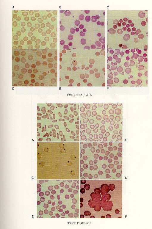
First Color Plate-upper
First Color Plate Lower
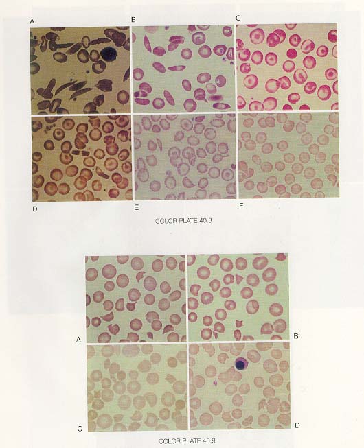
Second Color Plate upper
Second Color Plate Lower